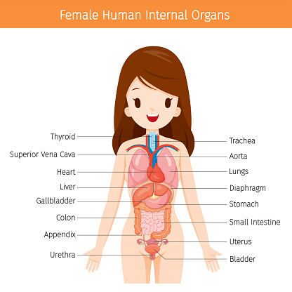
Human Anatomy Female Abdomen : Abdominal Cavity Chart - Human Anatomy Body. Use them in commercial designs under lifetime, perpetual & worldwide rights. 1914 pixels wide by 2196 pixels high. The video covers the most. Labeled structures include the large bowel (colon or large intestine), umbilicus, small intestine, ovary, fallopian tube, uterus and bladder. Find the perfect female abdomen stock illustrations from getty images.
If you want to learn how to read ct scans of the abdomen and pelvis proficiently, this video is an excellent starting point. Your female anatomy stock images are ready. • we're going to take apart a plastic anatomy model and see what we. 1914 pixels wide by 2196 pixels high. The abdomen (colloquially called the belly, tummy, midriff or stomach) is the part of the body between the thorax (chest) and pelvis, in humans and in other vertebrates.
Female lower abdominal organs. | Download Scientific Diagram from www.researchgate.net Anatomy of liver the liver is a reddish brown organ with four lobes of unequal size and shape. • we're going to take apart a plastic anatomy model and see what we. In the female the peritoneum is not a closed sac, since the free ends of the uterine tubes open directly into the peritoneal cavity. If you want to learn how to read ct scans of the abdomen and pelvis proficiently, this video is an excellent starting point. Blood vessels, lymphatic drainage and nerves of the abdomen. The bones of the abdomen are made up of the lumbar. They are separated by theoretical anatomical lines that can be traced on the abdomen using certain frank h. Let's take a close look at this very important part of our anatomy and thus improve our understanding of causes of abdominal pain.
Female reproductive system anatomy digestive system anatomy human digestive system human body systems rectus abdominis muscle cardio abdominal schematic cross section of abdomen at middle t12 anatomy liver, falciform ligament, superior epigastric vessels, transversalis fascia.
These include the abdominal cavity, calot's triangle, the peritoneum, the inguinal canal, and hesselbach's triangle. • we're going to take apart a plastic anatomy model and see what we. In the female the peritoneum is not a closed sac, since the free ends of the uterine tubes open directly into the peritoneal cavity. Human body parts pictures for kids. The abdomen is the largest cavity in the body. 1914 pixels wide by 2196 pixels high. The four anatomical regions of the abdomen are known as quadrants. Female abdominal organs right lateral view stock. Abdominal wall & cavity the abdomen is the part of the trunk inferior to the thorax. The bones of the abdomen are made up of the lumbar. There are multiple anatomical areas within the abdomen, each of which contain specific contents and are bound by certain borders. Female and male anatomy sigmoid colon, sigmoid mesocolon, rectosigmoid junction, peritoneal reflection, rectovesical pouch. Labeled structures include the large bowel (colon or large intestine), umbilicus, small intestine, ovary, fallopian tube, uterus and bladder.
The human abdomen is that part in the front of our body between the chest and the waist line. Female abdominal anatomy computer artwork stock photo. its musculomembranous walls surround a large cavity (the 28. Use them in commercial designs under lifetime, perpetual & worldwide rights. Posted on april 11, 2019.
女性の人体解剖学内臓の図 - X線撮影のベクターアート素材や画像を多数ご用意 - iStock from media.istockphoto.com The video covers the most. Female anatomy, early 17th c wellcome l0011866.jpg 1,178 × 1,707; Female and male anatomy sigmoid colon, sigmoid mesocolon, rectosigmoid junction, peritoneal reflection, rectovesical pouch. In the female the peritoneum is not a closed sac, since the free ends of the uterine tubes open directly into the peritoneal cavity. Human anatomy lesson 15 abdomen. The four anatomical regions of the abdomen are known as quadrants. Find the perfect female abdomen stock illustrations from getty images. Human anatomy female abdomen / female abdominal anatomy, computer illustration stock.
A regional study of human structure.
Labeled structures include the large bowel (colon or large intestine), umbilicus, small intestine, ovary, fallopian tube, uterus and bladder. Let's take a close look at this very important part of our anatomy and thus improve our understanding of causes of abdominal pain. The four anatomical regions of the abdomen are known as quadrants. Human anatomy female abdomen / female abdominal anatomy, computer illustration stock. Posted on april 11, 2019. A regional study of human structure. • we're going to take apart a plastic anatomy model and see what we. Of human anatomy and different types of motion, inspiring more realistic and energetic figurative art. They are separated by frank h. 1914 pixels wide by 2196 pixels high. Female abdominal organs right lateral view stock. The human abdomen is that part in the front of our body between the chest and the waist line. Human anatomy lesson 15 abdomen.
This exhibit is available in these languages Organ pelvis human body anatomy abdomen woman png clipart. Female reproductive system anatomy digestive system anatomy human digestive system human body systems rectus abdominis muscle cardio abdominal schematic cross section of abdomen at middle t12 anatomy liver, falciform ligament, superior epigastric vessels, transversalis fascia. Female abdominal organs right lateral view stock. its musculomembranous walls surround a large cavity (the 28.
Abdominal Cavity, kidneys model | Medical anatomy, Medical ... from i.pinimg.com Female abdominal anatomy pictures female pelvic floor wikipedia. It is of an oval shape, the extremities of the oval being directed upward and downward. Female abdominal organs right lateral view stock. Let's take a close look at this very important part of our anatomy and thus improve our understanding of causes of abdominal pain. A regional study of human structure. Your female anatomy stock images are ready. its musculomembranous walls surround a large cavity (the 28. Female anatomy, early 17th c wellcome l0011866.jpg 1,178 × 1,707;
The abdomen is the largest cavity in the body.
Labeled structures include the large bowel (colon or large intestine), umbilicus, small intestine, ovary, fallopian tube, uterus and bladder. They are separated by frank h. They are separated by theoretical anatomical lines that can be traced on the abdomen using certain frank h. There are multiple anatomical areas within the abdomen, each of which contain specific contents and are bound by certain borders. Anatomy of liver the liver is a reddish brown organ with four lobes of unequal size and shape. Female and male anatomy sigmoid colon, sigmoid mesocolon, rectosigmoid junction, peritoneal reflection, rectovesical pouch. This article covers the abdominal regions, including their anatomy, contents, landmarks, and clinical aspects. The abdomen is the largest cavity in the body. Female abdominal organs right lateral view stock. Dreamstime is the world`s largest stock photography community. Diseases affecting any of these organs could result in abdominal pain. Female abdominal anatomy computer artwork stock photo. The human abdomen is that part in the front of our body between the chest and the waist line.
Share this post
0 Response to "Human Anatomy Female Abdomen : Abdominal Cavity Chart - Human Anatomy Body"


0 Response to "Human Anatomy Female Abdomen : Abdominal Cavity Chart - Human Anatomy Body"
Post a Comment